Protein aggregation
Application range: protein aggregation
Particles size and shape characterization plays an important role in biotechnological formulations based on protein engineering.
An increasing amount of studies on proteins are appearing in bio-pharmaceutical research, and Occhio has developed two instruments for aggregation and quantification in protein suspensions. Average protein size is about 4 to 6nm. During production or conditioning and storage, the aggregation process may spontaneously begin. The protein aggregate has various size ranges from hundreds of nm to hundreds of µm.
To see testing for protein aggregation done by occhio (click here) and see Apn16-BT001!
These are our main objectives:
Detect all aggregates, from small to coarse
We integrated a large sensor with 12 Megapixels in combination with high resolution optics
Recognize transparent aggregate while deciphering single particles from cluster particles
We developed a special threshold that improves the detection of low contrast particles; Lenses and backlight are also chosen to improve the contrast.
Small sample quantity analysis
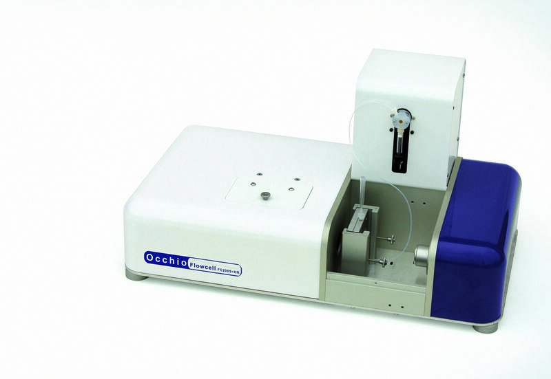
To maintain sample integrity, we minimized the fluid path in the flow cell and incorporated a small syringe to reduce the sample quantity required while maintaining unrivaled particle detection and characterization.
100% sample analysis
An advanced particle tracking function, combined with full cell image capture allows 100% sample coverage.
Easy cell replacement
The cell can be cleaned by water or other solutions. Due to frequent use of the instrument, it is necessary to replace the cell when cleaning is not possible. Occhio reduces the replacement cost of a single cell. Cell replacement is very easy, simply replace the microchip cell held in place by the support and a perfect seal is achieved automatically by closing the support and no tools are needed!
Determination of foreign particles
Complex filtering functions discriminate any foreign particles such as silicon, oil, or other external contamination.
Fully computer-controlled method includes motorized magnifications and autofocus procedure
All the steps of the analysis are controlled by a computer and the magnification is controlled by a precise, position cognizant motor. Five preset resolutions are possible just by specification in the SOP. A motorized focus scans the entire cell thickness and Callisto 3D software chooses the best focus position.
Instrument calibration
Calibration is independently conducted for each user’s method.
Callisto 3D software is fully compliant with CFR21 part 11 regulation
Multi sample dispenser
We reduced analysis time and labor costs by integrating the automatic dispenser robot with the instrument. Up to 96 samples per plate, and includes cleaning and tip replacement after each run.

Ipac 1
From 400nm to mm range, dispersion surface 140mm

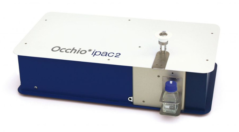
Ipac 2
From 400nm to mm range, dispersion surface 140mm
